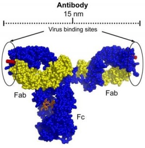
Before using antibodies for the detection of proteins by immunohistochemistry (IHC), all epitopes of the tissue sample must be blocked to prevent nonspecific antibody binding. Otherwise, the antibodies or other detection reagents may be connected to all the epitopes of the sample, regardless of the specificity.
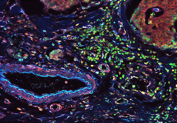
Fluorescent detection of vimentin and lamin IHC B1 in normal colon tissue. Sections of normal human colon cancer tissues were blocked with BSA buffer Thermo Scientific blocking windows before incubation with anti-vimentin antibodies and anti-lamin B1 antibody, followed by incubation with Thermo Scientific DyLight 405 goat anti-mouse or Thermo Scientific DyLight 549 goat anti-rabbit secondary antibodies, respectively. The cell nuclei were against green.
Introduction
In principle, any protein that does not have a binding affinity for the target or probe in the assay components can be used for blocking. In practice, however, some proteins work better than others, because they are easier to connect to non-specific sites (also known as reactive sites) and stabilize the function of other system components. In fact, any protein or protein mixture that works best for all IHC experiments and empirical testing is essential to get the best results for a given specific antibody and the support system combination.
General Procedures blocks
Blocking step for IHC is most often done after all other sample preparation and prior to sample incubation with the primary antibody. The general protocol be incubated with fixed, built, installed, and clears the sample IHC mask with appropriate blocking buffer for a period of time from 30 minutes to overnight at room temperature or at 4 ° C or on the basis protocol optimized for each specific antibody and antigen target. Enough after washing is essential to remove excess protein, which can interfere with the detection of the target antigen blocking step.
Types of blocking buffer
Normal serum
Normal serum is a common blocking agent, since serum contains antibodies that bind to the reactive sites, and thus prevent the nonspecific binding of the secondary antibody used in the assay. A key factor, however, is to use serum of the species in which the secondary antibody is generated, unlike the case of the primary antibody. The main types of serum antibody binds to the reactive sites, but these secondary antibodies recognize non-specific antibodies bound to antibodies bound to the target antigen.
The protein solutions
In addition, serum proteins, concentrated buffer in 0.1 to 5% bovine serum albumin (BSA), the skim milk powder or gelatin is frequently used to cover all the proteins present in the sample. Forcing this approach to overcome the primary antibodies for blocking the protein binding to cognate ligands, while reducing non-specific binding of anti-substance has a greater affinity for non-specific epitopes than do proteins buffering. Although these pads can be easily performed in the laboratory, for best results, they should be fresh before use, which increases the time and expense of color IHC.
Prefabricated commercial Buffers
Ready blocking buffers are available for securing a sample to antibody treatment. These buffers may contain high concentrations of single purified protein or protein free of proprietary compounds. One advantage of using commercial blockers is that there are many options available that work better than gelatin or casein and improved life.